It may be less than a millimetre long and just a day old, but there’s a lot going on in a fruit fly embryo as it tucks and folds itself into shape. Thin sheets of cells (epithelia) are on the move, wrapping themselves around the oval-shaped embryo and sealing together along its back.
This process, known as dorsal closure, works a bit like a suitcase being zippered shut. It’s an essential step in the embryo’s journey from fertilised egg to wriggling maggot and is a source of endless fascination to Jérôme Solon and his team at the CRG.
A few years ago, Solon made the important discovery that one of the driving factors of this biological zippering comes from the cells in the gap (known as the amnioserosa) that shrink down and die. The rest comes from cables of sturdy molecules known as actin and myosin – actomyosin – that connect the cells surrounding the gap on the embryo’s surface and pull them together.
But although Solon and his team had figured out how forces could be generated by these cells, there was another mystery still waiting to be solved.
Although these epithelial sheets are moving around all the time as their cells change in size, shape and number, the surface always stays smooth and taut, with no baggy wrinkles, gaps appearing between cells or breaks in the cell borders due to being over-stretched. This suggests that the cell shape and the tension in the tissue sheet is somehow being adjusted so it always stays the same. So what’s going on?
To find out, Solon’s team did a very simple experiment that nobody had done before: they took some fruit fly embryos and squished them very, very carefully between thin pieces of glass. Pressing down on the embryos stretches their epithelial covering, while releasing the pressure causes the tissue sheets to temporarily ruffle up before becoming smooth again.
“We realised that the answer must lie in the connections between the individual cells,” he says. These junctions are made from assemblies of the adhesive molecule cadherin, which form little chains supported by actomyosin filaments. Importantly, the junctions can quickly be lengthened or shortened by adding or removing molecules as required.
“Our idea was to squeeze and stretch embryos to see what happens to the cell junctions as they are pushed together or pulled apart,” says Solon. “This requires very careful precision, down to the level of a thousandth of a millimetre: squeeze too much and the embryo will burst, not enough and you don’t see anything.”
By carefully watching what was happening at the junctions as they squeezed or released the embryos, Solon and his team realised that the lengths of the cell connections were changing rapidly. Their results show that the tension between cells is maintained by quickly adjusting the junction length to accommodate the level of stretch.
“If the connection becomes too stretched then the cell brings in more actomyosin to pull everything back together again. And if it is too floppy and loose because the cells are bunching up, then some of the junction is removed so that the correct tension can be restored,” he explains.
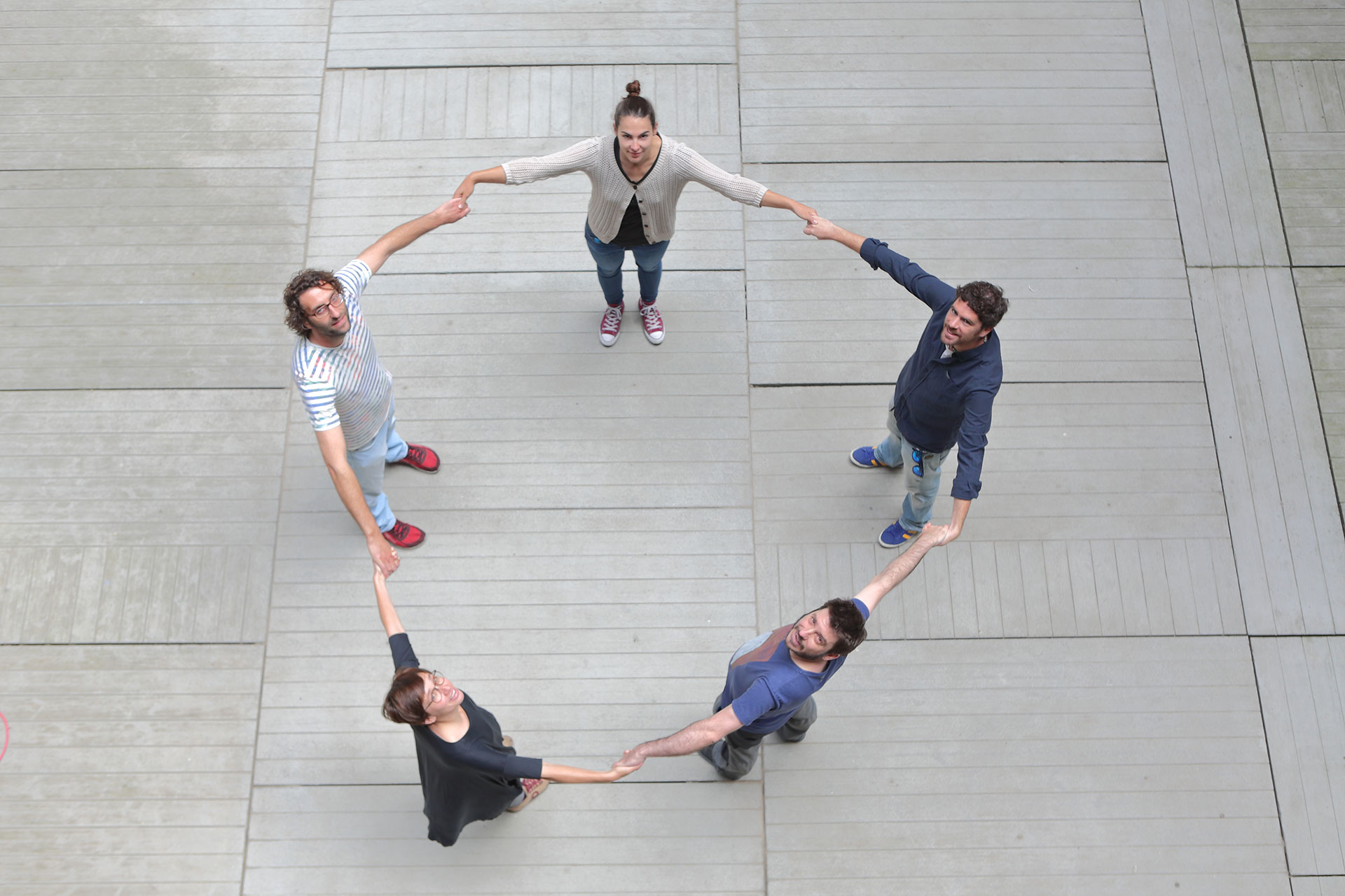
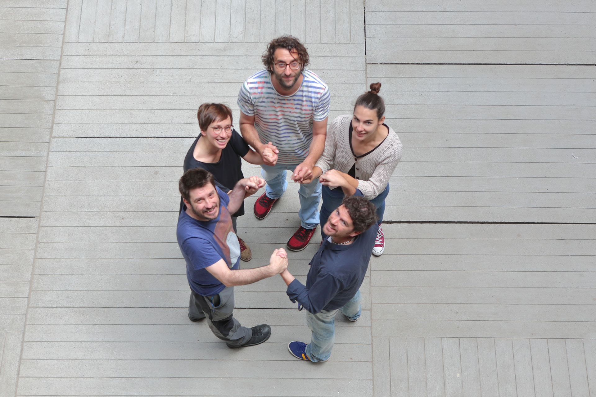
To understand how this works, imagine a chain of people holding hands across a shifting, stretchy ground, all trying to maintain a steady tension. If they drift too far apart and the chains risks being torn, they can call in some more friends and hold on tighter to keep the chain strong and sturdy. But if they start to bunch too closely together, then these extra people can drop out again.
“We were very excited to see the reaction of the cells – we weren’t expecting it to be that dramatic for such a simple experiment, but it is amazing how quickly the cells adapt as the embryo changes shape,” Solon says. “Our previous paper showed that cells shrink in size to help to close the embryo and now we have solved the mystery of how they make sure their shape and integrity is maintained while they do that, through constant readjustment of the junctions between them.”
Published in Developmental Cell, this discovery sheds light on how tissues maintain their structure through the dynamic shifts and changes that happen during development. Other researchers have discovered similar processes at work in epithelia in other organisms such as developing frog tadpoles, suggesting that they are a fundamental part of the mechanics of life.
“Embryos are so dynamic – it is amazing the things that go on in a matter of minutes, and we are always finding something we weren’t expecting about how cells are moving and responding to external pressures,” Solon says. “It’s an incredible self-organising system that can cope with all kinds of changes, yet still every embryo looks the same. I think that’s really impressive.”